Doubling the resolution of fluorescence-lifetime single-molecule localization microscopy with image scanning microscopy
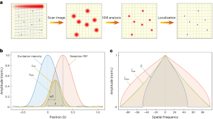
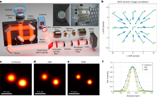
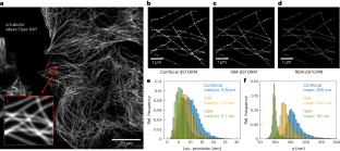
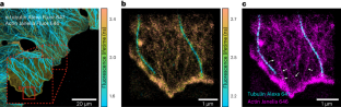
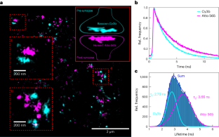
In this study, we integrate a single-photon detector array into a confocal laser scanning microscope, enabling the combination of fluorescence-lifetime single-molecule localization microscopy with image scanning microscopy. This unique combination delivers a twofold improvement in lateral localization accuracy for single-molecule localization microscopy (SMLM) and maintains its simplicity. Moreover, the addition of lifetime information from our confocal laser scanning microscope eliminates chromatic aberration, particularly crucial for achieving few-nanometre resolution in SMLM. Our approach, named fluorescence-lifetime image scanning microscopy SMLM, is demonstrated through direct stochastic optical reconstruction microscopy and DNA point accumulation for imaging in nanoscale topography experiments on fluorescently labelled cells, showcasing both resolution enhancement and fluorescence-lifetime multiplexing capabilities.
This is a preview of subscription content, access via your institution
Access options
Access Nature and 54 other Nature Portfolio journals
Get Nature+, our best-value online-access subscription
cancel any time
Subscribe to this journal
Receive 12 print issues and online access
206,07 € per year
only 17,17 € per issue
Buy this article
- Purchase on SpringerLink
- Instant access to full article PDF
Prices may be subject to local taxes which are calculated during checkout
Three-dimensional total-internal reflection fluorescence nanoscopy with nanometric axial resolution by photometric localization of single molecules
Article Open access 22 January 2021

Fast widefield scan provides tunable and uniform illumination optimizing super-resolution microscopy on large fields
Article Open access 24 May 2021
Optimizing imaging speed and excitation intensity for single-molecule localization microscopy
Article 17 August 2020
Data availability
Code availability
References
- Betzig, E. et al. Imaging intracellular fluorescent proteins at nanometer resolution. Science313, 1642–1645 (2006). ArticleADSGoogle Scholar
- Huang, B., Wang, W., Bates, M. & Zhuang, X. Three-dimensional super-resolution imaging by stochastic optical reconstruction microscopy. Science319, 810–813 (2008). ArticleADSGoogle Scholar
- Heilemann, M. et al. Subdiffraction-resolution fluorescence imaging with conventional fluorescent probes. Angew. Chem. Int. Ed.47, 6172–6176 (2008). ArticleGoogle Scholar
- Sharonov, A. & Hochstrasser, R. M. Wide-field subdiffraction imaging by accumulated binding of diffusing probes. Proc. Natl Acad. Sci. USA103, 18911–18916 (2006). ArticleADSGoogle Scholar
- Schnitzbauer, J., Strauss, M. T., Schlichthaerle, T., Schueder, F. & Jungmann, R. Super-resolution microscopy with DNA-PAINT. Nat. Protoc.12, 1198–1228 (2017). ArticleGoogle Scholar
- Reinhardt, S. C. et al. Ångström-resolution fluorescence microscopy. Nature617, 711–716 (2023). ArticleADSGoogle Scholar
- Thiele, J. C. et al. Confocal fluorescence-lifetime single-molecule localization microscopy. ACS Nano14, 14190–14200 (2020). ArticleGoogle Scholar
- Oleksiievets, N. et al. Single-molecule fluorescence lifetime imaging using wide-field and confocal-laser scanning microscopy: a comparative analysis. Nano Lett.22, 6454–6461 (2022). ArticleADSGoogle Scholar
- Chizhik, A. I., Rother, J., Gregor, I., Janshoff, A. & Enderlein, J. Metal-induced energy transfer for live cell nanoscopy. Nat. Photon.8, 124–127 (2014). ArticleADSGoogle Scholar
- Isbaner, S. et al. Axial colocalization of single molecules with nanometer accuracy using metal-induced energy transfer. Nano Lett.18, 2616–2622 (2018). ArticleADSGoogle Scholar
- Thiele, J. C. et al. Isotropic three-dimensional dual-color super-resolution microscopy with metal-induced energy transfer. Sci. Adv.8, 2506 (2022). ArticleGoogle Scholar
- Sheppard, C. R. Super-resolution in confocal imaging. Optik80, 53–54 (1988). Google Scholar
- Müller, C. B. & Enderlein, J. Image scanning microscopy. Phys. Rev. Lett.104, 198101 (2010). ArticleADSGoogle Scholar
- Sheppard, C. J., Mehta, S. B. & Heintzmann, R. Superresolution by image scanning microscopy using pixel reassignment. Opt. Lett.38, 2889–2892 (2013). ArticleADSGoogle Scholar
- Sheppard, C. J., Castello, M., Tortarolo, G., Vicidomini, G. & Diaspro, A. Image formation in image scanning microscopy, including the case of two-photon excitation. J. Opt. Soc. Am. A34, 1339–1350 (2017). ArticleADSGoogle Scholar
- Sheppard, C. J. et al. Pixel reassignment in image scanning microscopy: a re-evaluation. J. Opt. Soc. Am. A37, 154–162 (2020). ArticleADSGoogle Scholar
- Kner, P., Chhun, B. B., Griffis, E. R., Winoto, L. & Gustafsson, M. G. Super-resolution video microscopy of live cells by structured illumination. Nat. Methods6, 339–342 (2009). ArticleGoogle Scholar
- Reymond, L. et al. SIMPLE: structured illumination based point localization estimator with enhanced precision. Opt. Express27, 24578–24590 (2019). ArticleADSGoogle Scholar
- Reymond, L., Huser, T., Ruprecht, V. & Wieser, S. Modulation-enhanced localization microscopy. J. Phys.: Photonics2, 041001 (2020). ADSGoogle Scholar
- Cnossen, J. et al. Localization microscopy at doubled precision with patterned illumination. Nat. Methods17, 59–63 (2020). ArticleGoogle Scholar
- Sun, Y. et al. Modulated illumination localization microscopy-enabled sub-10 nm resolution. J. Innov. Opt. Health Sci.15, 2230004 (2022). ArticleGoogle Scholar
- Kalisvaart, D. et al. Precision in iterative modulation enhanced single-molecule localization microscopy. Biophys. J.121, 2279–2289 (2022). ArticleADSGoogle Scholar
- York, A. G. et al. Instant super-resolution imaging in live cells and embryos via analog image processing. Nat. Methods10, 1122–1126 (2013). ArticleGoogle Scholar
- Schulz, O. et al. Resolution doubling in fluorescence microscopy with confocal spinning-disk image scanning microscopy. Proc. Natl Acad. Sci. USA110, 21000–21005 (2013). ArticleADSGoogle Scholar
- Tenne, R. et al. Super-resolution enhancement by quantum image scanning microscopy. Nat. Photon.13, 116–122 (2019). ArticleADSGoogle Scholar
- Rossman, U. et al. Rapid quantum image scanning microscopy by joint sparse reconstruction. Optica6, 1290–1296 (2019). ArticleADSGoogle Scholar
- Castello, M. et al. A robust and versatile platform for image scanning microscopy enabling super-resolution flim. Nat. Methods16, 175–178 (2019). ArticleGoogle Scholar
- Buttafava, M. et al. SPAD-based asynchronous-readout array detectors for image-scanning microscopy. Optica7, 755–765 (2020). ArticleADSGoogle Scholar
- Koho, S. V. et al. Two-photon image-scanning microscopy with SPAD array and blind image reconstruction. Biomed. Opt. Express11, 2905–2924 (2020). ArticleGoogle Scholar
- Slenders, E. et al. Cooled SPAD array detector for low light-dose fluorescence laser scanning microscopy. Biophys. Rep.1, 100025 (2021). Google Scholar
- Roth, S., Sheppard, C. J., Wicker, K. & Heintzmann, R. Optical photon reassignment microscopy (OPRA). Opt. Nano.2, 5 (2013). ArticleGoogle Scholar
- Roth, S., Sheppard, C. J. & Heintzmann, R. Superconcentration of light: circumventing the classical limit to achievable irradiance. Opt. Lett.41, 2109–2112 (2016). ArticleADSGoogle Scholar
- Fazel, M. et al. Fluorescence microscopy: a statistics-optics perspective. Rev. Mod. Phys.96, 025003 (2024). ArticleADSGoogle Scholar
- Stallinga, S., Radmacher, N., Delon, A. & Enderlein, J. Optimal transfer functions for bandwidth-limited imaging. Phys. Rev. Research4, 023003 (2022). ArticleADSGoogle Scholar
- Smith, C. S. et al. Structured illumination microscopy with noise-controlled image reconstructions. Nat. Methods18, 821–828 (2021). ArticleGoogle Scholar
- Holden, S. J., Uphoff, S. & Kapanidis, A. N. DAOSTORM: an algorithm for high-density super-resolution microscopy. Nat. Methods8, 279–280 (2011). ArticleGoogle Scholar
- Ovesný, M., Křížek, P., Borkovec, J., Švindrych, Z. & Hagen, G. M. ThunderSTORM: a comprehensive ImageJ plug-in for PALM and STORM data analysis and super-resolution imaging. Bioinformatics30, 2389–2390 (2014). ArticleGoogle Scholar
- Stein, S. C. & Thiart, J. TrackNTrace: a simple and extendable open-source framework for developing single-molecule localization and tracking algorithms. Sci. Rep.6, 37947 (2016). ArticleADSGoogle Scholar
- Linde, S. Single-molecule localization microscopy analysis with ImageJ. J. Phys. D: Appl. Phys.52, 203002 (2019). ArticleADSGoogle Scholar
- Stein, S., Thiart, J., Thiele, J. C. & Radmacher, N. Single-molecule localization microscopy with image scanning microscopy. GitLabhttps://gitlab.gwdg.de/igregor/single-molecule-localization-microscopy-with-image-scanning-microscopy (2023).
- Rieger, B. & Stallinga, S. The lateral and axial localization uncertainty in super-resolution light microscopy. ChemPhysChem15, 664–670 (2014). ArticleGoogle Scholar
- Zwettler, F. et al. Molecular resolution imaging by post-labeling expansion single-molecule localization microscopy (ex-SMLM). Nat. Commun.11, 3388 (2020). ArticleADSGoogle Scholar
- Thiele, J. C., Nevskyi, O., Helmerich, D. A., Sauer, M. & Enderlein, J. Advanced data analysis for fluorescence-lifetime single-molecule localization microscopy. Front. Bioinform.1, 740281 (2021). ArticleGoogle Scholar
- Wegel, E. et al. Imaging cellular structures in super-resolution with SIM, STED and Localisation Microscopy: a practical comparison. Sci. Rep.6, 27290 (2016). ArticleADSGoogle Scholar
- Oleksiievets, N. et al. Fluorescence lifetime DNA-PAINT for multiplexed super-resolution imaging of cells. Commun. Biol.5, 38 (2022). ArticleGoogle Scholar
- Kalisvaart, D., Hung, S.-T. & Smith, C. S. Quantifying the minimum localization uncertainty of image scanning localization microscopy. Biophys. Rep.4, 100143 (2024).
- Dani, A., Huang, B., Bergan, J., Dulac, C. & Zhuang, X. Superresolution imaging of chemical synapses in the brain. Neuron68, 843–856 (2010). ArticleGoogle Scholar
- Truckenbrodt, S. et al. X10 expansion microscopy enables 25-nm resolution on conventional microscopes. EMBO Rep.19, 45836 (2018). ArticleGoogle Scholar
- Tortarolo, G. et al. Focus image scanning microscopy for sharp and gentle super-resolved microscopy. Nat. Commun.13, 7723 (2022). ArticleADSGoogle Scholar
- Slenders, E. & Vicidomini, G. ISM-FLUX: MINFLUX with an array detector. Phys. Rev. Research5, 023033 (2023). ArticleADSGoogle Scholar
Acknowledgements
N.R., J.E. and S.O.R. acknowledge financial support from the Bundesministerium für Bildung und Forschung (BMBF), Germany, via project NG-FLIM (project nos. 13N15327 and 13N15328). J.I.G. acknowledges financial support from the European Union’s Horizon 2021 research and innovation programme under the Marie Skłodowska-Curie grant agreement no. 101062508 (project name: SOADOPP). J.E. and J.I.G. acknowledge financial support from the DFG through Germany’s Excellence Strategy EXC 2067/1-390729940. J.E. and O.N. thank the European Research Council (ERC) for financial support via project ‘smMIET’ (grant agreement no. 884488) under the European Union’s Horizon 2020 research and innovation programme. We thank A. Chizhik for providing us with the illustration of our ISM microscope setup, as well as for carefully reading the manuscript.
Author information
- These authors contributed equally: Niels Radmacher, Oleksii Nevskyi, José Ignacio Gallea.
Authors and Affiliations
- Third Institute of Physics—Biophysics, Georg August University, Göttingen, Germany Niels Radmacher, Oleksii Nevskyi, José Ignacio Gallea, Ingo Gregor & Jörg Enderlein
- Department of Chemistry, University of Oxford, Oxford, UK Jan Christoph Thiele
- Department of Neuro- and Sensory Physiology, University Medical Center Göttingen, Göttingen, Germany Silvio O. Rizzoli
- Cluster of Excellence ‘Multiscale Bioimaging: from Molecular Machines to Networks of Excitable Cells’ (MBExC), Universitätsmedizin Göttingen, Göttingen, Germany Silvio O. Rizzoli & Jörg Enderlein
- Niels Radmacher
